Mon–Fri 8:00 - 18:00
YouTube

Learn more

Good to know

  • A macular hole causes distortion and a spot in central vision; an epiretinal membrane may produce similar signs.
  • OCT confirms the diagnosis. Surgery (vitrectomy) is discussed according to the difficulties experienced and the stage.

Our doctors

See also all our doctors and our publications.

In the event of sudden loss of vision or pain, see eye emergencies.

Macular Hole and Epiretinal Membrane: When Central Vision Is Affected

Macular holes and epiretinal membranes are two conditions affecting the macula, the central area of the retina responsible for precise, detailed vision. Although distinct, these conditions can reduce visual acuity and distort images, affecting reading, recognising faces and daily activities.

Macular Hole: A Gap in the Centre of Vision

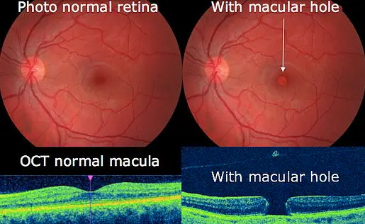
A macular hole is an opening in the centre of the macula, like a small gap in this fragile tissue. It generally occurs with age, when the vitreous (the transparent gel filling the eye) separates from the retina and exerts excessive traction on the macula.

Comparison of a normal macula and a macular hole

Different types of macular holes:

  • Full-thickness macular hole : The hole extends through all layers of the retina.
  • Lamellar hole (or macular pseudohole) : Only the superficial layers of the retina are affected.

Symptoms:

  • Reduced central visual acuity
  • Distorted vision (metamorphopsia): straight lines appear wavy or curved
  • A dark or blurred spot in the centre of the visual field

Epiretinal Membrane (ERM): A Veil over the Macula

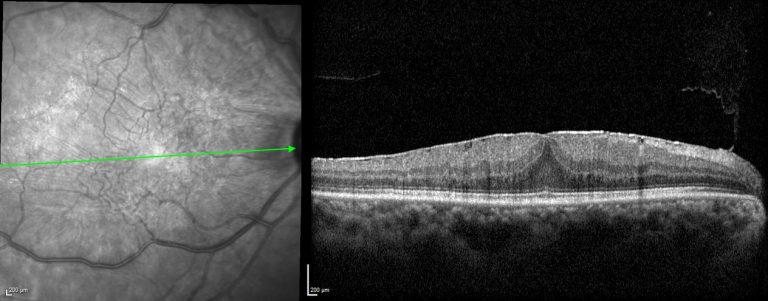
Also called a “macular pucker”, an epiretinal membrane is a thin membrane of scar tissue forming on the surface of the macula. It may contract and wrinkle the retina, disrupting central vision.

Epiretinal membrane: photograph and OCT cross-section

Other terms for ERM:

  • Idiopathic retinal membrane
  • Cellophane maculopathy
  • Preretinal fibrosis

Symptoms:

  • Blurred and distorted vision (metamorphopsia)
  • Reduced visual acuity, especially when reading and doing detailed work
  • Seeing images as smaller or larger than they really are (micropsia or macropsia)

Care at the Centre Médical des Aravis

The Centre Médical des Aravis has expertise in diagnosing and treating macular holes and epiretinal membranes.

Diagnosis:

  • Fundus examination
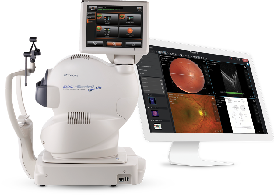
  • OCT (Optical Coherence Tomography) : provides detailed images of the different layers of the retina

Treatment:

Treatment depends on the severity of the condition and its impact on vision. It may be:

  • Monitoring : for mild, non-progressive cases
  • Surgery : vitrectomy to remove the vitreous and the membrane, followed by a gas injection to flatten the retina

Our experienced ophthalmic surgeons have mastered the most advanced micro-invasive techniques to optimise the chances of visual recovery and minimise complications.

If you experience problems with central vision, please consult our specialists at the Centre Médical des Aravis.

Topcon Maestro2 OCT device and its imaging interface
A question about your situation?

Your ophthalmologist answers your questions during the consultation. For a sudden symptom or sudden loss of vision, seek advice promptly: see the eye emergencies page.

Book an appointmentContact receptionOur locations

Our practices

Annecy-Argonay

645 route de Champ Farçon, 74370 Argonay

Faverges

27 place Gambetta, 74210 Faverges

Rumilly

5 rue Frédéric Girod, 74150 Rumilly

Sallanches

35 rue Emma Lanche, 74700 Sallanches

04 50 23 03 23Book an appointmentDirections and parkingReception Monday to Friday, 8 am – 6 pm