Mon–Fri 8:00 - 18:00
YouTube

Your centre's equipment

Discover the equipment used for eye tests and ocular imaging at the Centre médical des Aravis.

BY LOCATION

Our equipment, centre by centre

Argonay · Centre médical des Aravis

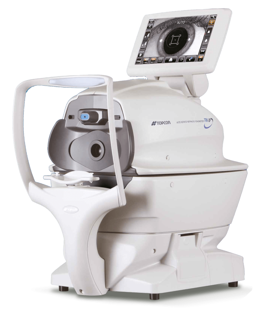
  • Topcon TRK-2P autorefractorAutomatic refraction measurement, the first stage of the vision assessment.
  • Slit lamps with photographyMicroscopic examination of the eye’s structures, with photographs for the medical record.
  • YAG laserLaser used for certain treatments, such as clouding that develops after cataract surgery.
  • PASCAL laserLaser for retinal photocoagulation.
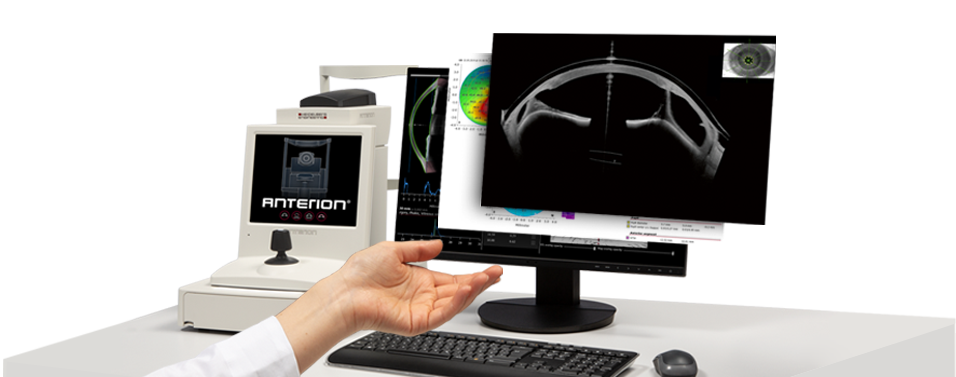
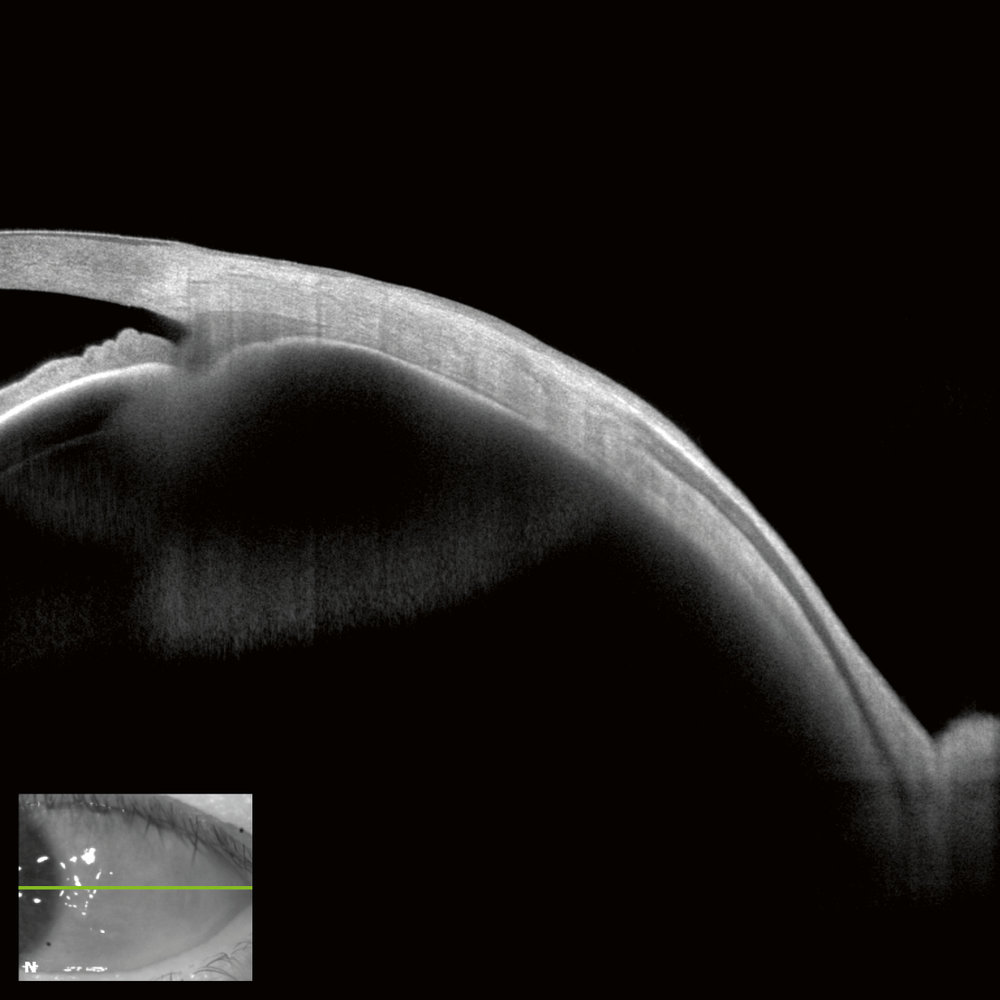
  • OCT ANTERIONCross-sectional imaging of the front of the eye: cornea, anterior chamber, lens.
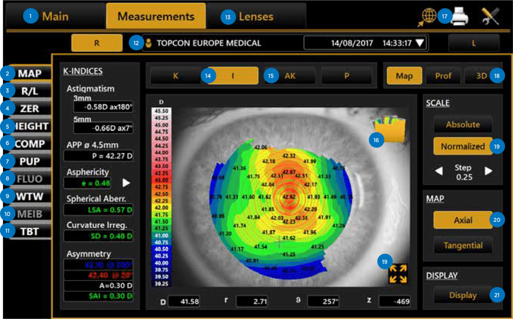
  • CA-800 corneal topographerMapping of corneal curvature.
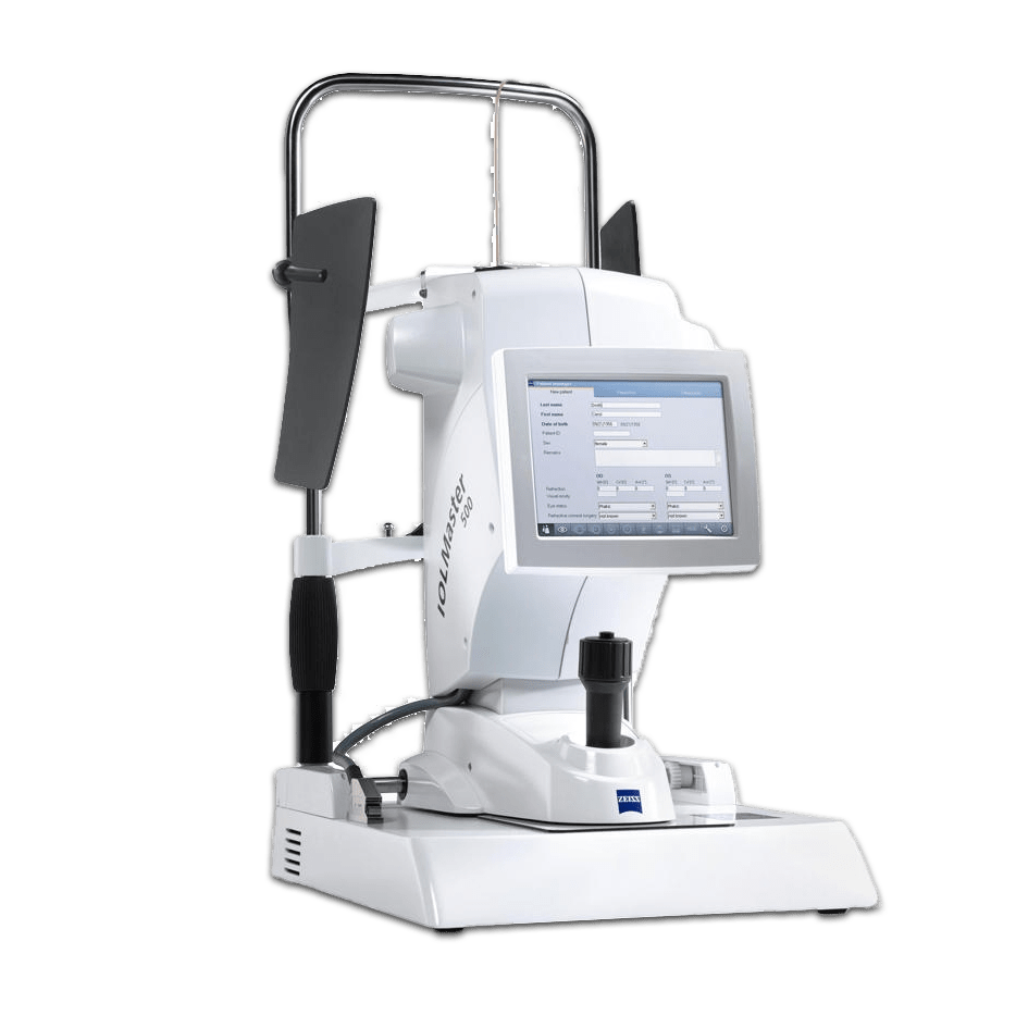
  • IOLMaster 700Optical biometry: eye measurements used to calculate the cataract lens implant.
  • Nidek specular microscopeExamination and counting of corneal endothelial cells.
  • Ultrasound biometerMeasurement of eye length using ultrasound.
  • Heidelberg OCT angiography systemCross-sectional imaging of the retina and its blood vessels, without an injection.
  • OCT Maestro 2Cross-sectional imaging of the retina and optic nerve.
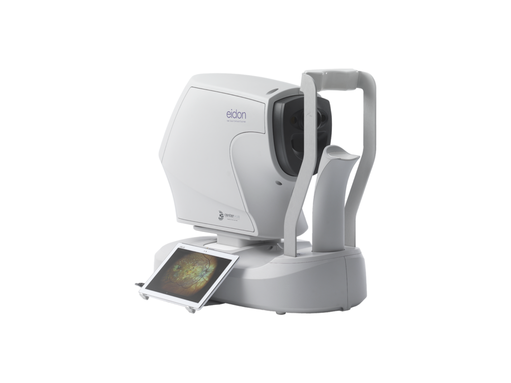
  • EIDON wide-field fundus cameraWide-field photographs of the fundus.

Annecy Laser Vision · Argonay

  • Bausch + Lomb Teneo 2 excimer laserRefractive surgery laser.
  • Johnson & Johnson Vision Elita femtosecond laserFemtosecond laser for refractive surgery.

Faverges

  • OCT Maestro 2Cross-sectional imaging of the retina and optic nerve.
  • YAG laserLaser used for certain treatments, such as clouding that develops after cataract surgery.

Rumilly

  • OCT Maestro 2Cross-sectional imaging of the retina and optic nerve.

Your ophthalmologist chooses examinations according to the reason for your consultation. Details of several examinations are provided below.

Our equipment

Your ophthalmologist chooses examinations according to the reason for your consultation and your follow-up needs.

This page presents the centre's equipment and its role in examining vision and the structures of the eye. Not every test is needed at every consultation.

Digital illustration of an eye and its analysis

Autorefractors

The autorefractor measures refraction by sending a beam of light through the patient's eye and measuring how much the light is deflected as it passes through the eye.

Le résultat de la mesure est utilisé pour déterminer la prescription de lunettes et de lentilles nécessaire pour corriger votre vision.

The refractor also measures values for keratometry (bulging of the cornea), of pachymetry (corneal thickness) and tonometry (tonus oculaire ou pression oculaire).

Topcon TRK-2P autorefractor and tonometer
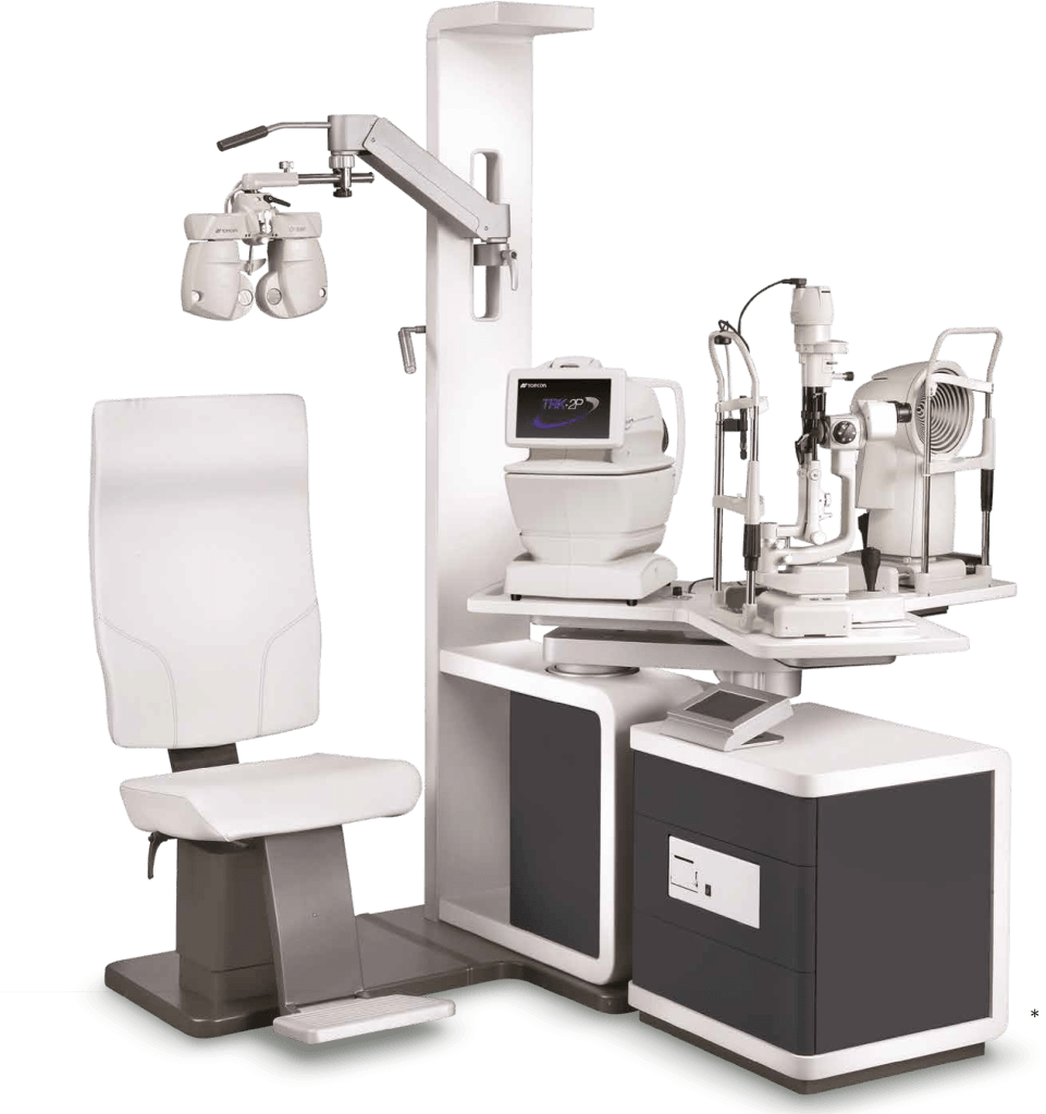
Topcon ophthalmic consultation table and unit

Examination table

Le bilan visuel et l'examen ophtalmologique sont réalisés grâce à une table d'examen dédiée à l'ophtalmologie. Celle-ci est composée du autorefractor, keratometer, pachymeter, tonometer, of a phoropter permettant de tester la vision.

Un microscope (ou lampe à fente) permettra l'examen des structures oculaires : paupières, conjonctive, globe oculaire.

Focimeter

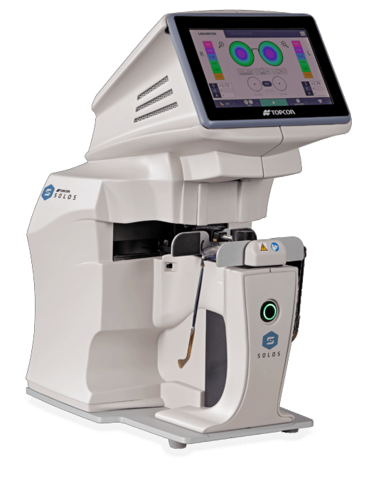
The focimeter measures the parameters of spectacle lenses.

Topcon lensmeter for measuring spectacle lenses

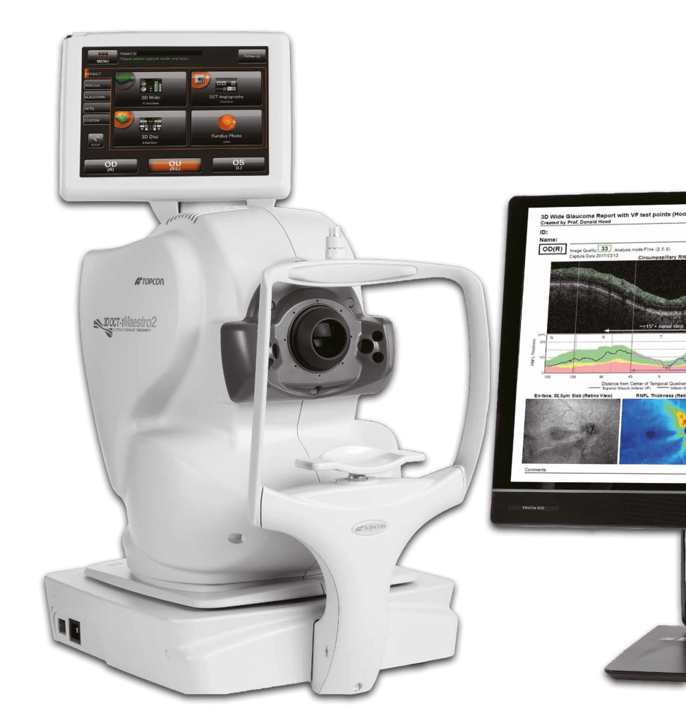
OCT Maestro 2

Un OCT (sigle de l'anglais Optical Coherence Tomography, ou Tomographie en Cohérence Optique en français) est un appareil médical utilisé pour obtenir des images en coupe transversale de la retina, of the choroid of the eye and the optic nerve. It is mainly used by ophthalmologists to diagnose and monitor various eye conditions, such as age-related macular degeneration (AMD), the diabetic retinopathy and the glaucoma.

L'OCT fonctionne en envoyant un faisceau de lumière à travers l'œil du patient et en mesurant la quantité de lumière qui est reflétée ou absorbée par les différentes couches de tissu de l'œil. Les données recueillies sont utilisées pour créer une image en coupe transversale de l'œil qui peut être examinée par un médecin pour détecter des anomalies ou des changements dans les tissus oculaires.

OCT is a non-invasive and painless method pour obtenir des images de l'œil.

Topcon OCT device
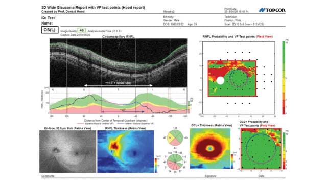
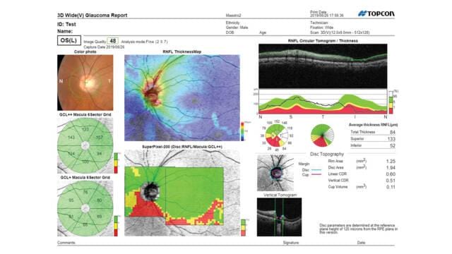
OCT analysis: retinal cross-sections and thickness maps
OCT analysis of the retina and optic nerve
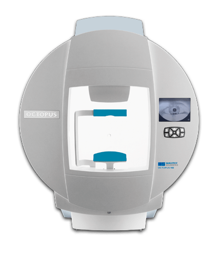
Haag-Streit Octopus perimeter for visual field testing

Visual field

Le champ visuel est la zone de l'espace que l'on peut voir sans bouger les yeux. Il comprend tout ce que vous voyez devant vous lorsque vous regardez droit devant vous, ainsi que les parties que vous pouvez voir en regardant vers les côtés sans bouger votre tête.

The visual field is important because it allows us to detect objects and movements in our immediate surroundings, which is essential for survival. It is also used to assess eye health and diagnose certain eye conditions.

Il existe plusieurs tests pour mesurer le champ visuel, tels que le test de confrontation, le test de Goldmann et le test de campimétrie automatisé. Ces tests permettent de déterminer si le champ visuel est normal ou s'il y a des limitations ou des anomalies, et peuvent être utilisés pour diagnostiquer et suivre des pathologies telles que le glaucome, la dégénérescence maculaire et d'autres troubles de la vision.

The Octopus 900 offers all commonly used standard static tests (automated perimetry). For central field testing, there are physiology-based G patterns following the retinal nerve fibres, and 32, 30-2 and 24-2 patterns. For the macula, there are the physiology-based M pattern and the 10-2 pattern. With the rapid TOP strategy, full threshold testing can be completed in just 2 to 4 minutes. The commonly used Esterman binocular test, a neurological pattern and a peripheral glaucoma pattern are also included. A Goldmann visual field test can also be performed with this device.

Ocular biometry

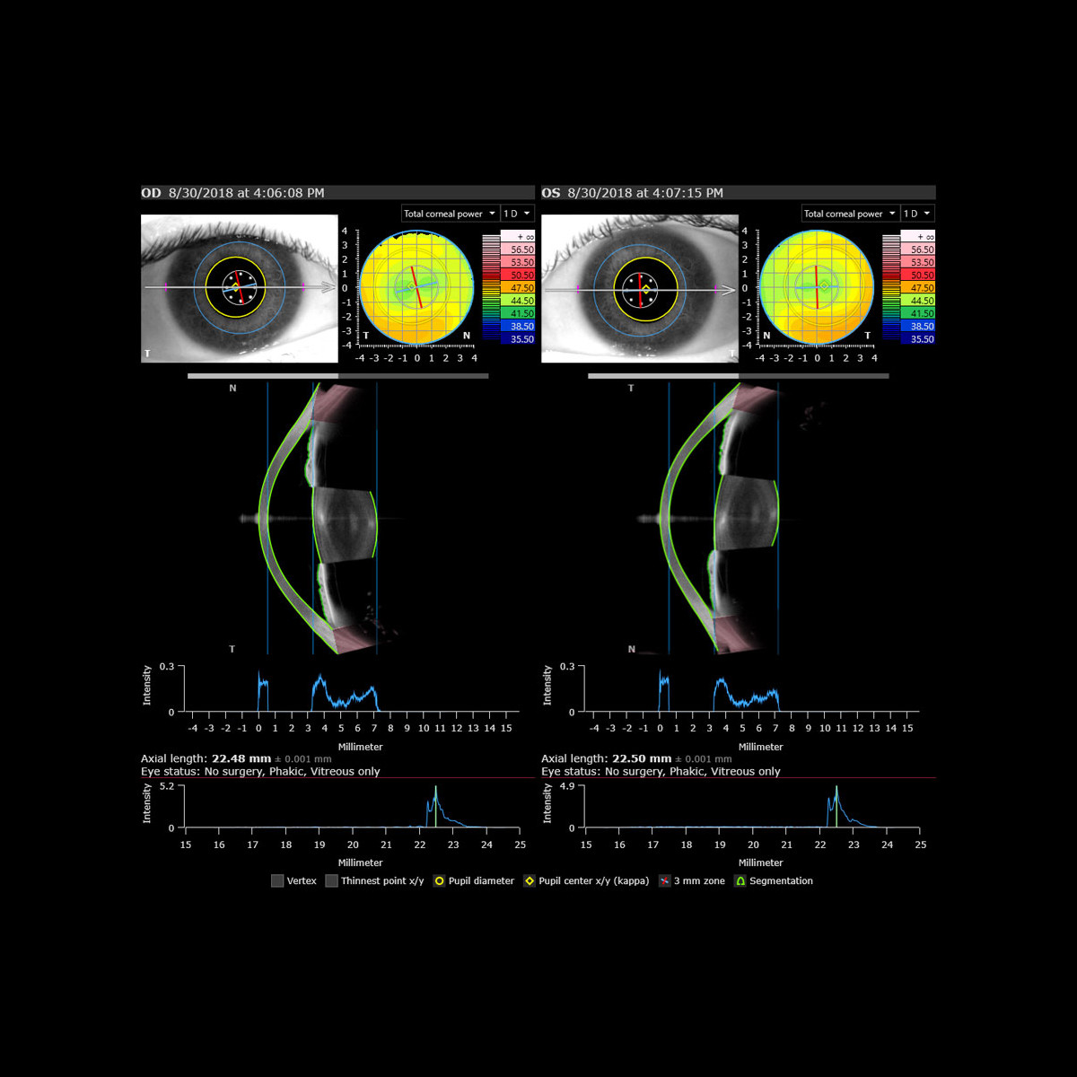
Infrared biometers work by sending a beam of light through the eye and measuring the amount of light reflected.

The measurement result is used to determine the length of the eye, the curvature of the cornea and the size of the anterior chamber. These measurements allow the power of the cataract lens implant to be placed in the eye after cataract surgery to be calculated

Zeiss optical biometer

Corneal topographer

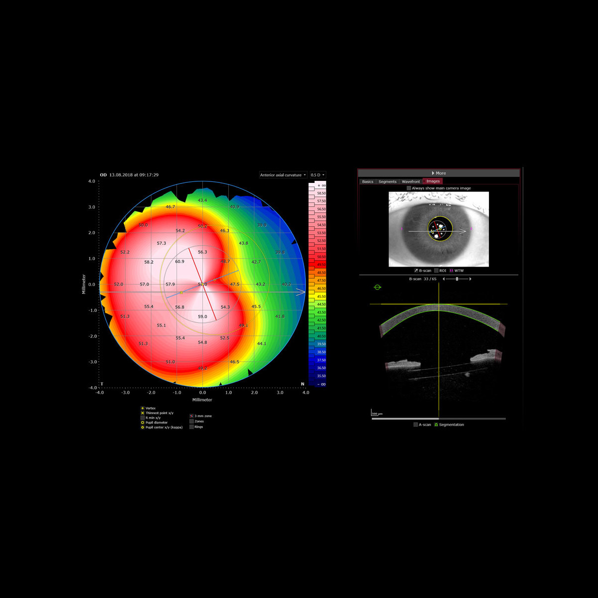
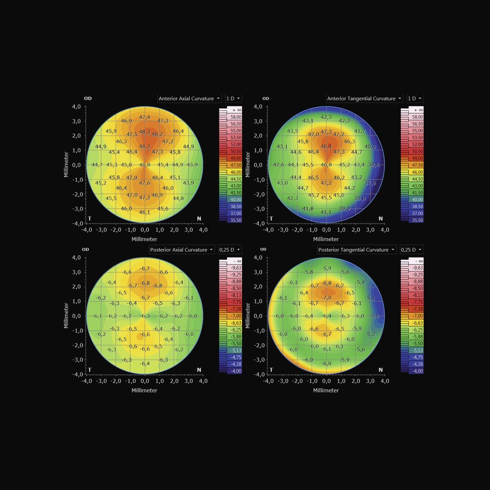
A corneal topographer is a device used to measure corneal curvature, the transparent curved part at the front of the eye that focuses light on the retina. It can be used to diagnose and monitor various eye conditions.

Le résultat de la mesure est utilisé pour créer une carte de la courbure de la cornée, appelée "topographie cornéenne", qui peut être examinée par un médecin pour détecter des anomalies ou des asymétries dans la courbure de la cornée. Les topographes cornéens sont considérés comme une méthode précise et fiable pour mesurer la courbure de la cornée et peuvent être utilisés pour aider à déterminer la prescription de lentilles de contact ou d'implants de chirurgie de cataracte nécessaire pour corriger la vision de l'individu.

Les topographes sont également utiles dans le bilan préalable à une chirurgie réfractive.

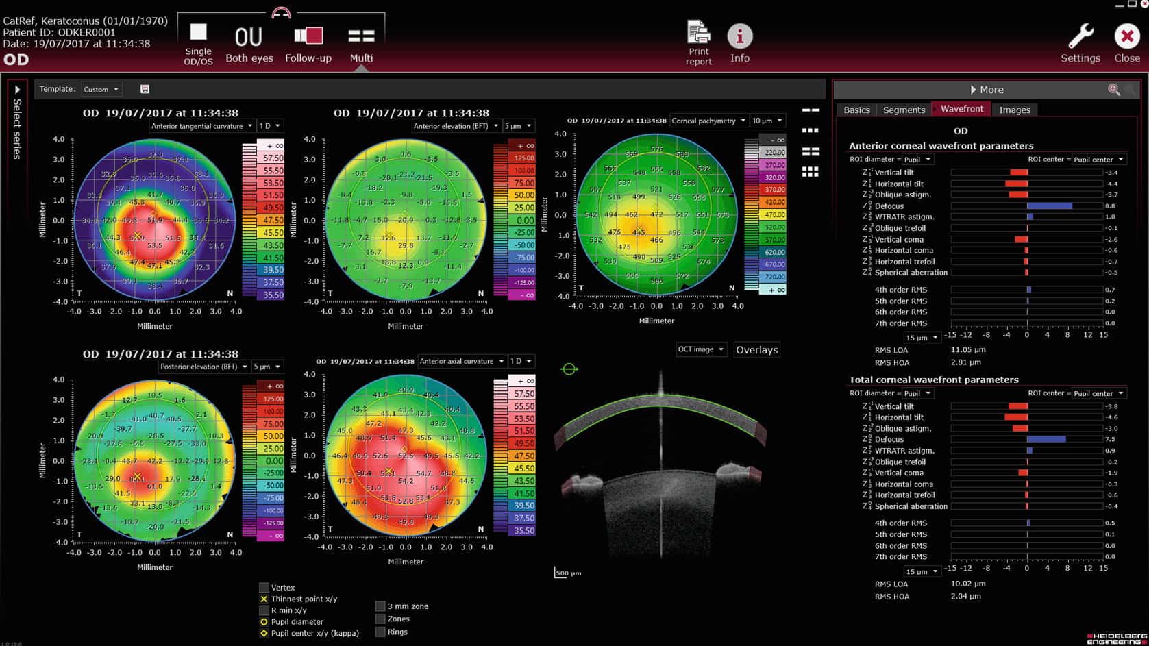
Corneal topography: colour map of the cornea
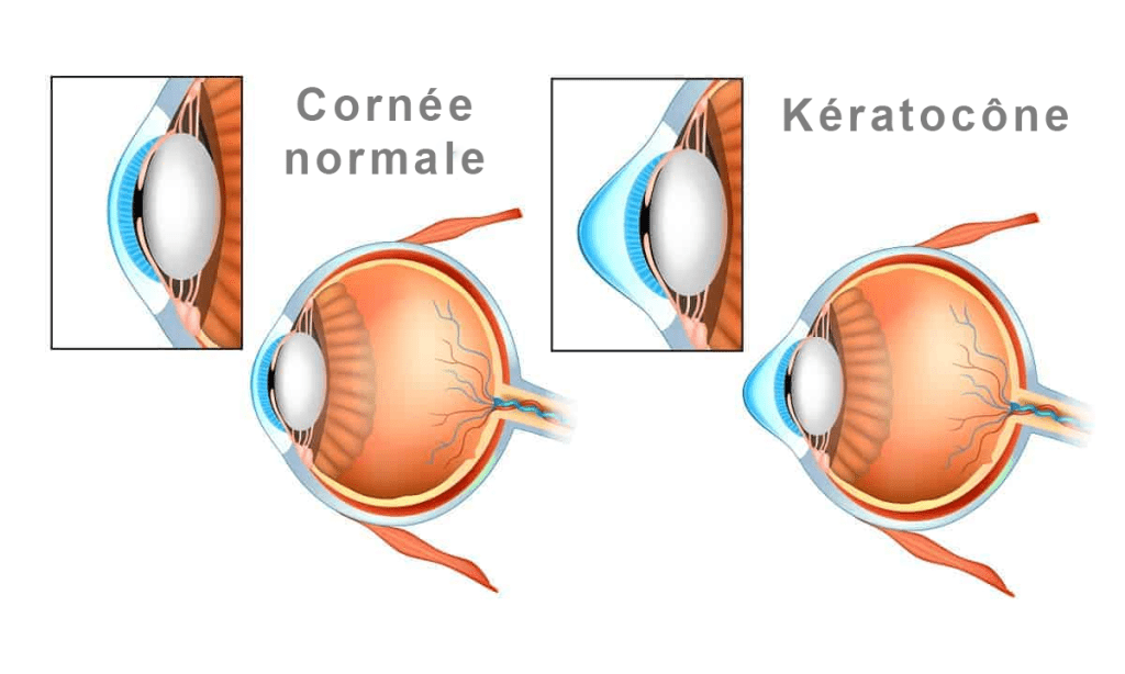
Three-dimensional comparison of a normal cornea and keratoconus

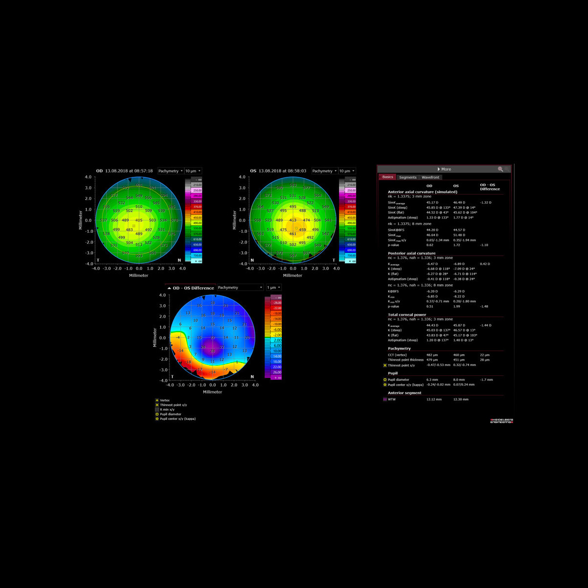
The CA-800 topographer and OCT Anterion topographer will be used in screening and monitoring keratoconus. Various measurements and scores are produced to help detect this progressive corneal condition as early as possible.

A corneal analysis of optical aberrations permet de déterminer les insuffisances optiques et aider à la planification d'une chirurgie réfractive ou de cataracte.

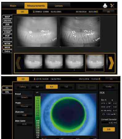
D'autres modules particulièrement intéressant sont disponibles avec le CA-800 :

- Pupillometry : Allowing the position and diameter of the pupil to be measured. This information is useful for Ortho-K treatment and refractive surgery.

- Fluorometry : enabling fluorescein images and videos for precise contact lens fitting. Tear quality, corneal condition and tear film break-up time can also be assessed.

- Meibomian gland analysis : useful for diagnosing and monitoring the treatment of meibomian gland dysfunction (posterior blepharitis).

- Simulation of soft and rigid contact lens fitting

Corneal topography: analysis screens and maps

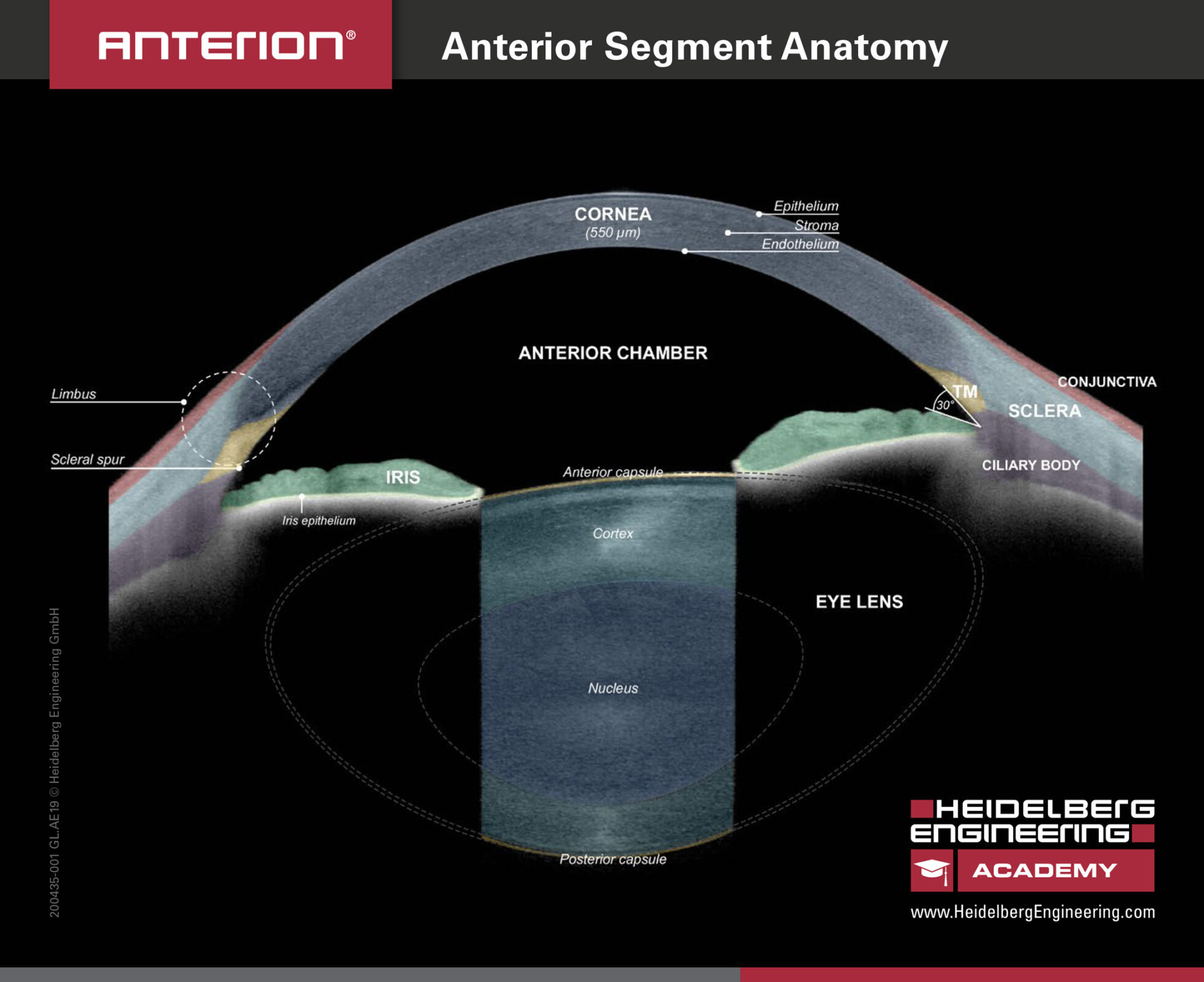
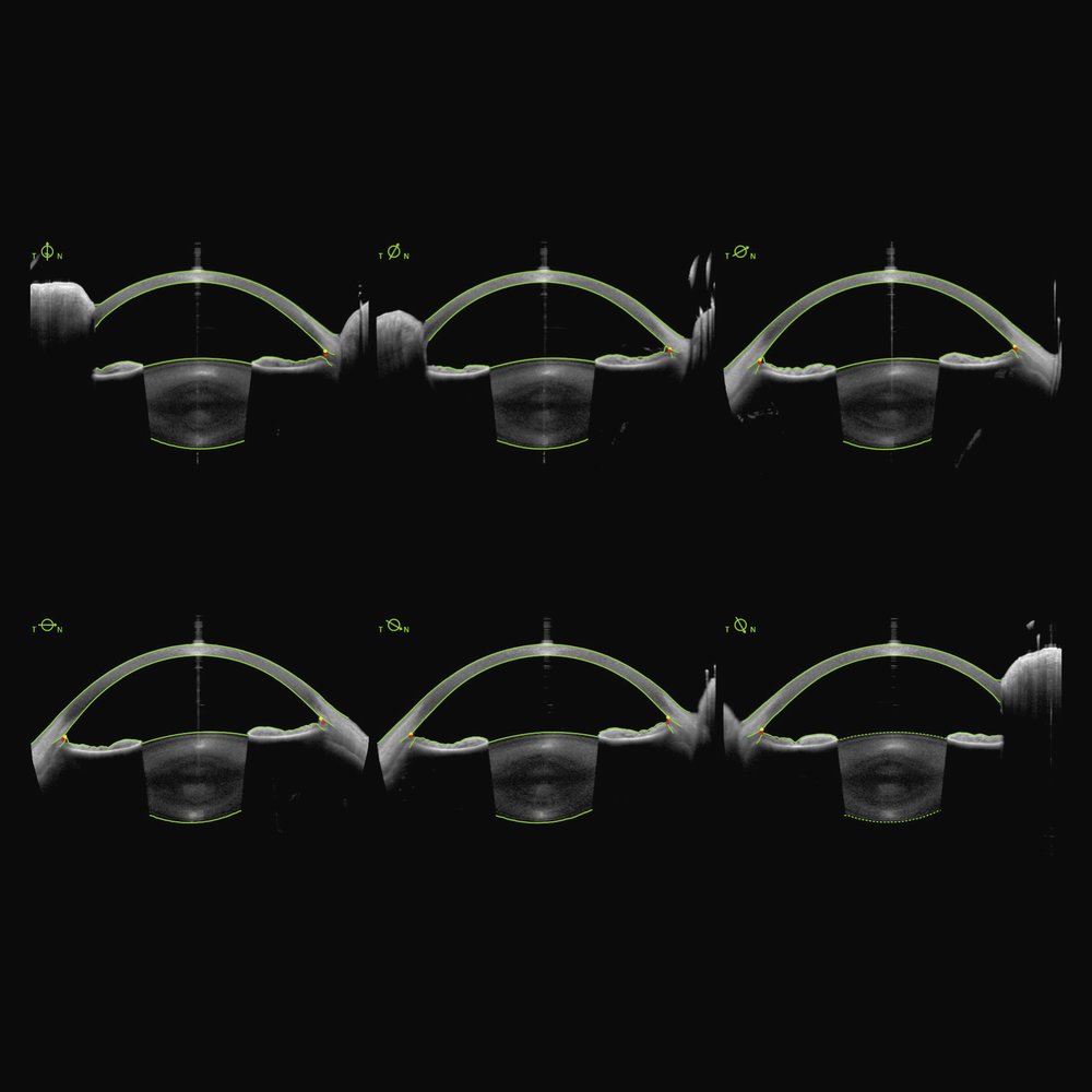
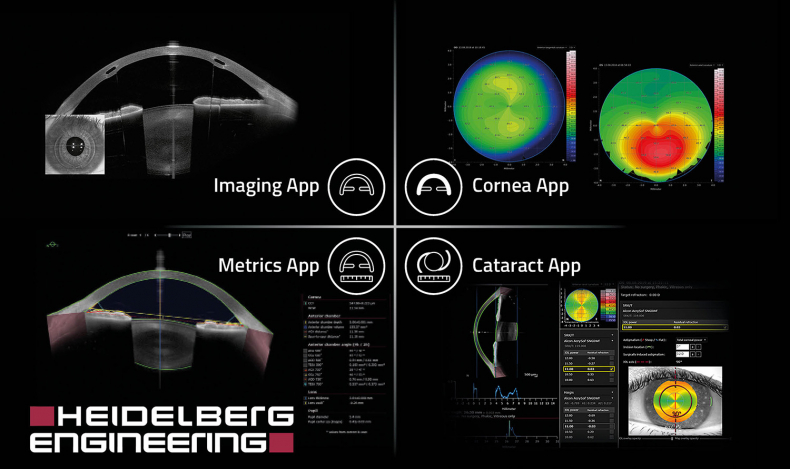
L'Anterion est une plateforme d'imagerie de dernière génération conçue pour offrir des résultats précis et convaincants dans l'évaluation de la structure de l'œil. Grâce à sa technologie OCT swept source, elle combine plusieurs applications en un seul appareil, permettant d'obtenir des mesures précises et d'identifier les pathologies de l'œil.

- Corneal topography : The Anterion maps the outer structure of the eye, allowing the practitioner to assess the front and back curvature of the cornea, as well as its thickness. This examination is essential before any refractive surgery to identify possible corneal contraindications.

- Biometry : The device provides precise preoperative measurements of artificial lens power before cataract surgery, helping to select the most suitable implant.

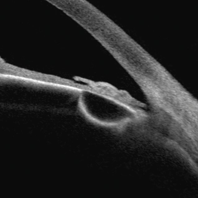
- High-definition imaging : Grâce à l'imagerie haute résolution, l'Anterion fournit des images et des mesures détaillées de tout le segment antérieur de l'œil, contribuant à un diagnostic plus fiable et à un suivi optimal.

Eidon retinal camera

Non-mydriatic retinal camera

Le centre d'ophtalmologie des Aravis est équipé de l'iCare EIDON, un système d'imagerie rétinienne TrueColor à large champ de vision. Cet appareil combine les avantages de la Scanning Laser Ophtalmoscopy (SLO) avec les caractéristiques de l'imagerie rétinienne classique pour établir de nouvelles normes en matière de performance.

- Advanced imaging technologies : The iCare EIDON offers various imaging modes, including TrueColor, blue, red and red-free images, as well as infrared confocal images. These modes provide a precise and faithful view of the retina. Ultra-high-resolution imaging: The iCare EIDON provides high-quality imaging, even in the presence of cataracts or media opacities, without needing to dilate the pupils (minimum diameter of 2.5 mm).

- Automated operation : The device can operate in fully automatic or manual mode, allowing a smooth and quick examination through its auto-alignment, autofocus, auto-exposure and auto-capture features.

- Wide-field visualisation : The device offers a field of view of 60° to 160° in a single image, which can be extended to 200° using the Ultra-Widefield module available at the centre des Aravis. This allows examination of both the central retina and its periphery, improving the detection and monitoring of peripheral conditions.

L'EIDON est un outil précieux pour le diagnostic et la surveillance des maladies rétiniennes, offrant une qualité d'image exceptionnelle et une interface conviviale.

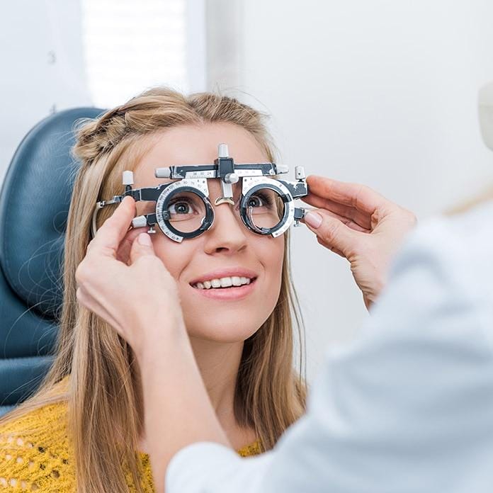
Patient examined with a trial frame

Our objectives

  • Individualised approach
  • Listening
  • Complementary roles of professionals

Preparing for your visit

Find practice addresses and ways to contact our team.

Our practices

Annecy-Argonay

645 route de Champ Farçon, 74370 Argonay

Faverges

27 place Gambetta, 74210 Faverges

Rumilly

5 rue Frédéric Girod, 74150 Rumilly

Sallanches

35 rue Emma Lanche, 74700 Sallanches

04 50 23 03 23Book an appointmentDirections and parkingReception Monday to Friday, 8 am – 6 pm