Dry Eye Updates (2025): From Diagnosis to Treatment Innovations – Summary of Dr Florian Baudin’s Presentation
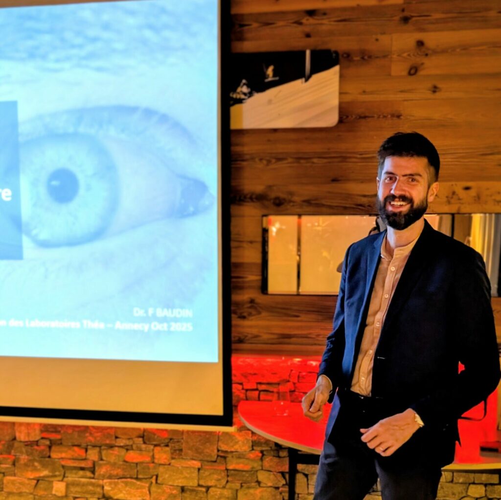
Dr Florian BAUDIN, an ophthalmologist at Centre Médical des Aravis, recently led a presentation for ophthalmologists in Haute-Savoie, invited by Théa Pharma laboratories, on the latest advances in the management of dry eye disease. This condition, a true “modern epidemic”, affects up to 20% of the population and significantly impacts quality of life. This article provides a comprehensive summary of the key points covered, from updated pathophysiological mechanisms to the most recent therapeutic innovations, integrating TFOS DEWS III recommendations.
To Better Understand: What is Dry Eye?
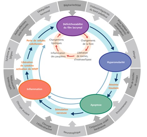
Dry eye is not simply a lack of tears. It is a complex disease in which the delicate balance of the eye’s surface is disrupted. Imagine a very thin protective film on the surface of your eye: the tear film. When this film becomes unstable or evaporates too quickly, the eye is no longer properly protected.
This triggers what is called a “vicious cycle”:
- Tear film instability: The film “breaks” too quickly between blinks.
- Hyperosmolarity: The tears become too concentrated (“too salty”).
- Inflammation: This abnormal concentration irritates the surface of the eye and triggers inflammation.
- Damage: The inflammation damages the cells on the surface of the eye.
- Worsening: This damage makes the tear film even more unstable… and the cycle continues.
The goal of modern treatments is to break this vicious cycle to restore a healthy balance to the eye’s surface.
Physiopathology: Understanding the Mechanisms at Play 🔬
Instability (TBUT < 10s) exposes the epithelium. The resulting hyperosmolarity is a central pathogenic factor, activating pro-inflammatory pathways (MAPK, NF-κB) and inducing cell apoptosis. It is a driving force, not just a consequence.
Inflammation is fundamental. It involves the release of cytokines (IL-1β, TNF-α) and MMP-9, which degrade the corneal barrier and lead to the loss of goblet cells. MGD (Meibomian Gland Dysfunction) is the most common cause of dry eye, with its own vicious cycle (obstruction, inflammation). The forms are often mixed.
The TFOS DEWS II has incorporated neuro-sensory abnormalities. Dry eye can be a chronic pain syndrome (“pain without stain”), sometimes associated with systemic diseases. The study of the ocular microbiome is a new frontier; dysbiosis could contribute to chronic inflammation.
TFOS DEWS III (2025): A New Era for Management 💡
Le rapport TFOS DEWS III, publié en 2025, marque un changement de paradigme majeur :
- New Definition: Emphasizes the “symptomatic” nature of the disease.
- Simplified Diagnosis: Based on a validated questionnaire (OSDI-6 ≥ 4) AND at least one positive clinical sign (NIBUT < 10s, Osmolarity ≥ 308 mOsm/L, or abnormal surface staining).
- Abandon of the “Evaporative vs. Aqueous” Classification: Considered suboptimal.
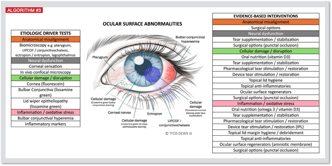
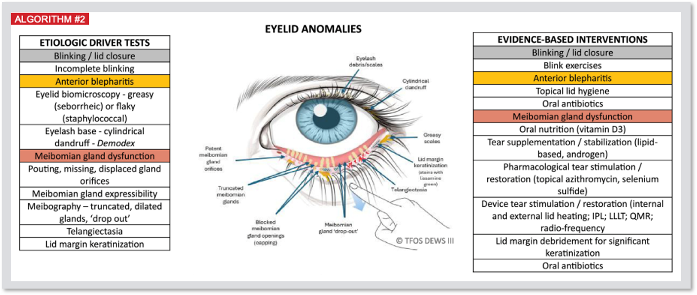
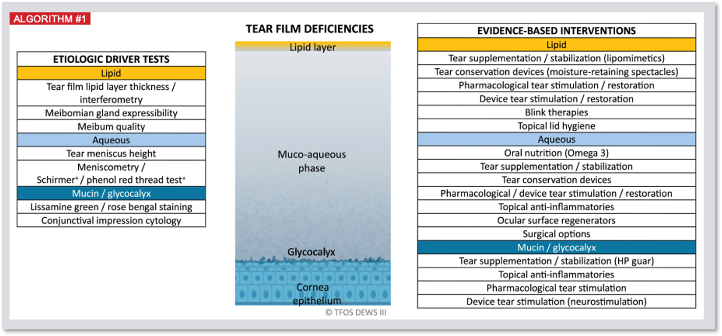
- New Approach Based on “Etiological Drivers”: Identifying specific triggers for each patient, classified into 3 domains:
- Deficiencies in the Tear Film (lipid, aqueous, mucin/glycocalyx)
- Eyelid Abnormalities (blinking, free edge, DGM, Demodex)
- Ocular Surface Abnormalities (anatomy, nerve, cells, inflammation)
- 3 Targeted Treatment Algorithms: Replace the step-by-step approach, linking “drivers” to evidence-based therapies for personalized and multimodal medicine.
Modern Diagnosis: Tools and Biomarkers 📈
- Subjective Assessment
- Examen Clinique Objectif
- Advanced Tools
OSDI-6, a simplified version of the OSDI, is recommended for screening (positive score ≥ 4) and follow-up. The SPEED questionnaire is also a relevant option.
Thorough examination under a slit lamp: eyelids, eyelashes, blinking, lacrimal meniscus. Measurement of tear film stability: TBUT (< 10s pathological), ideally using a non-invasive method (NIBUT). Vital stains: Fluorescein (KPS, Oxford score) and Lissamine Green (vitalized conjunctival cells).
Meibography: Infrared visualization of Meibomian glands, essential for diagnosing DGM and dropout.
Tear Osmolarity: Key biomarker (> 308 mOsm/L or ODG difference > 8 mOsm/L), measurable in the clinic.
Integrated Platforms (e.g., LacryDiag): Non-invasive analysis combining NIBUT, meibography, interferometry, and meniscusometry for a comprehensive assessment.
Therapeutic Strategies: A Personalized Approach 💊
The treatment remains progressive, but now adapts to the identified “drivers.” The goal is to restore homeostasis.
Artificial tears are the first line of treatment. Prioritize formulations without preservatives (BAK is toxic). Modern formulations target specific mechanisms.
Focus on Théalose® (3% Trehalose + 0.15% HA): Hypotonic solution without preservatives. Trehalose acts as an osmoprotectant, protecting cells from hyperosmolar stress, while hyaluronic acid hydrates and lubricates. It targets a key driver in the vicious cycle. Proven efficacy against HA alone (superiority on OSDI and TBUT) and against Systane® (superiority in symptoms). Modulates the inflammatory response.
Necessary when lubricants are insufficient.
Topical Corticosteroids: Effective for flares (“flares”), but long-term use is limited (side effects IOP/cataract). Choice of molecule and preservative-free status are crucial.
Focus Softacort® (Hydrocortisone 3.35mg/ml preservative-free): Low-potency corticosteroid in single-dose format. Minimises risk of IOP elevation. Efficacy demonstrated on hyperaemia, OSDI, TBUT without IOP variation. Treatment of choice for mild/moderate flares, especially in glaucoma-risk patients.
Immunomodulators (Maintenance treatment): The Ciclosporine is the reference. Inhibits T-lymphocyte activation, blocks pro-inflammatory cytokines, restores tear production. Delayed effect (several months).
Focus Ciclograft® (Ciclosporine 2%): High concentration, used for very severe dry eye and post-transplant cases via Compassionate Access (hospital prescription). Dr Baudin’s personal data on 55 patients show notable efficacy (78% major/moderate improvement) in refractory cases, with good general tolerance (12-month study).
Focus Vevizye® (Ciclosporine 0.1% water-free – EyeSol®): New formulation improving bioavailability, tolerance and residence time. Rapid action demonstrated on signs: significant improvement in corneal staining from Day 15 (57% responders). Good tolerance. Ideal if significant keratitis is present.
For severe, resistant, or cases with significant dry eye syndrome (DES).
Physical Treatments for DES:
– Intense Pulsed Light (IPL): Multifactorial action (photothermolysis of telangiectasias, antimicrobial/Demodex effect, photobiomodulation, thermal effect liquefying meibum). Proven efficacy (case reports, meta-analyses). Standard protocol: 3–4 sessions spaced 2–4 weeks apart. Duration per session: approximately 10–15 minutes.
– Thermal Pulsation (e.g., LipiFlow®): Internal heat + external pressure.
– Plasma Energy (Jett Plasma): Emerging technology (ionised gas using DC current). Targets hyperkeratinisation and the free edge biofilm via reversible electroporation and cell stimulation. Protocol: 1 to 3 sessions spaced 4–6 weeks apart, depending on severity. Duration: 5–10 minutes per eyelid. Initial promising results on OSDI, TBUT, and meibography. Positioned in the “Eyelids” algorithm of the TFOS DEWS III.
Autologous Blood Therapies:
– Autologous Serum (AS): Mimics tear composition (growth factors, vitamins).
– Platelet-Rich Plasma (PRP) / Growth Factors (PRGF): Higher concentration of growth factors.
Scleral Lenses: Alternative for very severe dry eye. Creates a tear reservoir, protects the cornea, and neutralises irregularities. Improves vision, comfort, and quality of life. A specific examination is performed to perfectly adapt the lens to the eye.
Ciclograft® in the Treatment of Severe Dry Eye Syndrome: Analysis on 55 Patients Treated in Our Center
La sécheresse oculaire est une pathologie fréquente, mais certaines formes sévères et réfractaires aux traitements conventionnels représentent un véritable défi thérapeutique. Dans ces cas complexes, des options plus puissantes sont nécessaires pour contrôler l’inflammation chronique et préserver la surface oculaire. Une étude rétrospective a été menée sur l’utilisation de la 2% Cyclosporine (Ciclograft®), une formulation hautement concentrée, chez nos patients présentant des pathologies inflammatoires sévères de la surface oculaire.Study Methodology
Cette analyse a porté sur une cohorte de 55 patients (of whom 63.6% were female), with an average age of 52.6 years. The indications for treatment with Ciclograft® were varied and represented complex cases:- Gougerot-Sjögren Syndrome (27.3%)
- Corneal Graft Rejection (21.8%)
- Atopic Keratoconjunctivitis (9.1%)
- Graft-versus-Host Disease (GVHD) (7.3%)
- Severe Ocular Rosacea (7.3%)
Main Results
Clinical Efficacy
Le traitement a montré une Major to moderate efficacy in 78.2% of patients. Dans ce groupe, 21,7% ont connu une amélioration qualifiée de “majeure”. Seuls 4,3% des patients n’ont montré aucune efficacité. Ces résultats sont particulièrement notables compte tenu de la sévérité et de la nature réfractaire des pathologies traitées.Improvement in Visual Acuity
Une amélioration statistiquement significative de l’acuité visuelle moyenne a été observée, passant de 0,42 à 0,49 (p=0,005) à la fin du suivi. Cela suggère que le contrôle de l’inflammation de surface a un impact direct et positif sur la fonction visuelle.Tolerance and Safety
Le traitement a été généralement bien toléré. 27,3% des patients ont rapporté des effets indésirables, qui étaient tous locaux (irritations, sensations de brûlure) et transitoires, conformes au profil attendu du produit. Le traitement a été interrompu chez 9,1% des patients en raison de ces effets locaux.Discussion and Limitations
Cette étude met en lumière l’efficacité notable de la ciclosporine à haute concentration pour des pathologies sévères, souvent en impasse thérapeutique. La bonne tolérance générale en fait une option stratégique précieuse pour gérer l’inflammation chronique. Il est cependant important de noter les limites de cette analyse : son caractère Retrospective, l’absence d’un groupe contrôle et un taux de perdus de vue relativement élevé (21,8%), qui limitent la portée des conclusions. Néanmoins, ces résultats fournissent des indications précieuses sur la place de ce traitement dans l’arsenal thérapeutique ophtalmologique.Conclusion
Pour les patients souffrant de sécheresse oculaire sévère et d’autres maladies inflammatoires de la surface oculaire ne répondant pas aux traitements standards, le Ciclograft® 2% appears to be an effective and safe therapeutic optionIt not only allows for significant control of inflammation, but also improves visual function, making it a valuable solution for the most complex cases.The Surgical Context: The Importance of Pre-Operative Care 👁️🗨️
Ophthalmic surgery (cataract, refractive surgery, glaucoma) is a major cause of iatrogenic dry eye. Mechanisms include inflammation, sectioning of corneal nerves, drug toxicity (preservatives such as BAK, MMC for glaucoma), and eyelid speculum trauma.
Impact on Cataract Surgery: The tear film is the first refractive surface. Its instability distorts biometry (keratometry), leading to implant calculation errors (up to 0.5D) and refractive surprises. This impact is amplified with “premium” implants (multifocal, toric), which degrade vision quality (halos, contrast) and create the “10/10 Unhappy Patient”. A stable ocular surface is non-negotiable for these surgeries, aiming for perfect vision not only in quantity but also in quality.
Essential Perioperative Management:
- Pre-operative: Systematically screen for dry eye (e.g., using the ASCRS algorithm with the SPEED II questionnaire). Postpone surgery if the dry eye is “visually significant” (OSDI > 22, TBUT < 7s). Aggressively optimise the surface (lubricants, anti-inflammatories, DGM) before biometric measurements, ideally over several weeks.
- Intra-operative: Minimise desiccation (humidification) and phototoxicity.
- Post-operative: Prioritise eye drops without preservatives (post-operative and for dry eye). Manage residual inflammation (e.g., Softacort®).
The Therapeutic Horizon: New Molecules and Mechanisms 🚀
A change in approach: stimulating natural tear production.
Acoltremon (TRYPTYR eye drops): An agonist of the corneal cold-sensing receptor TRPM8. Approved by the FDA in May 2025.
Varenicline (Tyrvaya nasal spray): A nicotinic receptor agonist that stimulates the trigeminal nerve through the nose. Approved by the FDA in 2021. It restores all 3 layers of the tear film.
Perfluorohexyloctane (Miebo/EvoTears – Eye Drops): 100% non-aqueous, forms a single lipid layer to prevent evaporation. Targets the DGM. Approved by the FDA in May 2023.
Lifitegrast (Xiidra – Eye Drops): LFA-1 antagonist, blocks the interaction of T lymphocytes. A new anti-inflammatory pathway. Approved by the FDA in 2016.
Promising molecules:
PL9643: Melanocortin receptor agonist (resolves inflammation).
BRM421: Regenerative peptide derived from PEDF (stimulates limbal stem cells).
Reproxalap: Reactive Aldehyde Suppressing Protein (RASP) inhibitor, acting upstream of inflammation.
Research into dual-mechanism therapies.
Clinical Case: A Concrete Example of Improvement 🌟
Dr Baudin illustrated the effectiveness of the new strategies with a representative clinical case:
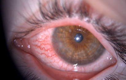
Initial Situation: 5-year-old girl with persistent redness of the right eye (OD) for over a month, accompanied by decreased vision and visual fatigue.
Symptoms: Intense burning sensation, visual fatigue in the morning. Blepharitis and clinical signs of ocular rosacea. Inflammation of the corneal limbus, more pronounced in the right eye (OD). Appearance of a sterile infiltrate and corneal neovascularisation.
Visual Acuity Without Correction:
Visual acuity OD = 9/10
Visual acuity OG = 10/10
Binocular visual acuity = 9/10
Final Refraction:
OD: +2.00 (-0.50 at 100°) = 6/10
OG: +2.50 (-0.75 at 85°) = 8/10
Binocular visual acuity = 9/10
Diagnosis:Severe infantile ocular rosacea with limbal insufficiency and blepharitis.
Initial Strategy: Frequent use of preservative-free lubricants + warm compresses + eyelid hygiene. Immediate introduction of a course of “gentle” corticosteroids without preservatives (Softacort® single-dose, 3 times/day) to manage the severe inflammatory flare.
Essential Refractive Care
- A cycloplegic refraction (Skiacol®) reveals a significant bilateral hypermetropia that was undiagnosed, which is the major cause of asthenopia.
- Prescription of a COT (corneal optical therapy) using glasses.
Observation: Image of the eye at diagnosis.
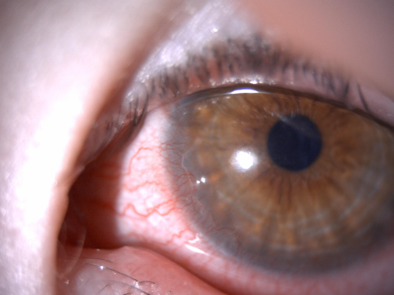
Improvement at Day 30: Théalose and Softacort show rapid improvement.
Progression: Significant reduction in burning. Improved overall comfort. However, upon discontinuation of corticosteroids: immediate recurrence.
Comment: Controlling hyperosmolarity and acute inflammation was crucial.
Strategy: In case of immediate relapse upon stopping corticosteroids, initiate long-term treatment with Ciclograft® 2% to control underlying chronic inflammation.
Observation: Progressive increase in optical correction to reduce the significant component attributable to eye strain.
Final Refraction: SKIA 2023
OD: +3.50 (-0.50 at 105°) = 10/10
OG: +2.75 = 9/10
Binocular VA = 10/10
Final refraction: SKIA 2024
OD: +4.00 (-1.00 at 110°) = 9/10
OG: +4.75 (-1.50 at 40°) = 9/10
AB = 9/10
Final refraction: SKIA 2025
OD= +4.75 (-1.50 at 120°)= 10/10
OG= +5.00 (-1.25 at 45°)= 10/10
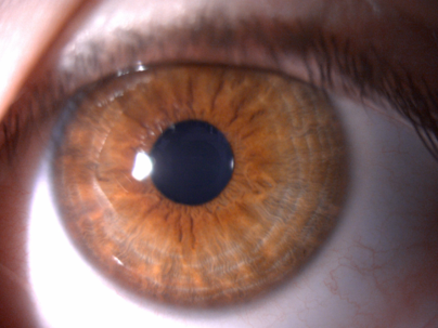
Complete resolution after 2 years of treatment: Ciclograft was well tolerated in conjunction with the introduction of Softacort
18-Month Evolution
- Rapid Improvement in surface condition following the introduction of the combined treatment (Softacort® followed by Ciclograft®).
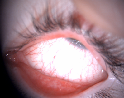
- Complete regression of the pannus neovascular, infiltrate and keratopathy lipidic within a few months.
- Allows for gradual weaning followed by complete discontinuation of cyclosporine in June 2025, without recurrence.
Final Result (October 2025)
- Visual acuity of 10/10 in each eye with corrective lenses.
- Calm ocular surface, with complete and inactive regression of corneal neovessels.
Commentary: The combination of immunomodulatory, immunosuppressive, and physical therapy for DGM is highly effective in severe cases.
Strategy: The entire treatment regimen has been discontinued. Continued use of full corrective lenses.
Observation: Image of the eye at 2 years.
General Conclusion: Personalized and Proactive Management ✅
The ocular surface disorder is a disease of homeostasis, with the vicious cycle as its central model. Diagnosis must be multimodal (OSDI-6, NIBUT, Osmolarity, Meibography). Treatment, guided by the “drivers” of TFOS DEWS III, becomes personalised and integrates therapeutic innovations as well as Artificial Intelligence.
Proposed Integrated Algorithm 2025:
- Mild Dry Eye: Education, preservative-free lubricants (Théalose® if hyperosmolarity is suspected/measured, or other artificial tear).
- Moderate Dry Eye: Foundation with Théalose® (or other artificial tear). Short course of Softacort® if flare-up (“flare”). Introduce an immunomodulator (e.g., Vevizye®, Ciclograft®) if chronic inflammation/keratitis, capitalizing on its rapid action on the signs. Consider IPL/Jett Plasma if DGM is predominant.
- Severe Dry Eye: Aggressive combined approach (Théalose®, Softacort® as induction, followed by cyclosporine). Discuss autologous serum, procedural treatments, scleral lenses.
Optimizing the ocular surface is key to therapeutic success, particularly in the context of surgery (cataract, refractive).
Scientific References 📚
References
Introduction & Pathophysiology
- Craig JP, et al. TFOS DEWS II Definition and Classification Report. Ocul Surf. 2017;15(3):276-283.
- Baudouin C, et al. The vicious circle of dry eye disease: a focus on the pathophysiology of meibomian gland dysfunction. Br J Ophthalmol. 2016;100(8):1050-1057.
- Bron AJ, et al. TFOS DEWS II pathophysiology report. Ocul Surf. 2017;15(3):438-510.
- Baudouin C. The pathophysiology of dry eye disease: a focus on the vicious circle of inflammation. Prog Retin Eye Res. 2013;36:270-282.
- AAO 2024: New study links dry eye to increased risk of psychiatric disorders and chronic pain syndromes. Ophthalmology Times. Accessed September 21, 2025.
TFOS DEWS III
- Wolffsohn JS, et al. TFOS DEWS III: A new era in the understanding and management of dry eye disease. Am J Ophthalmol. 2025 (In Press).
- Jones L, et al. TFOS DEWS III Management and Therapy Report. Am J Ophthalmol. 2025 (In Press).
Diagnosis
- Wolffsohn JS, et al. TFOS DEWS II Diagnostic Methodology report. Ocul Surf. 2017;15(3):539-574.
- Lemp MA, et al. The definition and classification of dry eye disease: report of the Definition and Classification Subcommittee of the International Dry Eye WorkShop (2007). Ocul Surf. 2007;5(2):75-92. (Note : L’article de 2011 est une extension de ces travaux).
Therapeutic Strategies
- Perumal N, et al. Combination Therapy with Trehalose and Hyaluronic Acid Restores Tear Lipid Layer Functionality by Ameliorating Inflammatory Response Protein Markers on the Ocular Surface of Dry Eye Patients. J Clin Med. 2025.
- HAS, Opinion of the National Commission for the Evaluation of Medical Devices and Health Technologies (CNEDIMTS) for THEALOSE, 18 June 2019.
- Kallab M, et al. Topical Low Dose Preservative-Free Hydrocortisone Reduces Signs and Symptoms in Patients with Dry Eye Disease. Adv Ther. 2020.
- Kuzmanović B, et al. A Retrospective Data Review Confirms That Topical Preservative-Free Hydrocortisone (Softacort®) Is Effective and Safe in the Treatment of Dry Eye Disease. Clin Ophthalmol. 2020.
- HAS, Opinion of the Transparency Commission for CICLOGRAFT (ciclosporine), 2023.
- Akpek E, et al. Pooled results from two pivotal randomized controlled clinical trials (ESSENCE-1 and ESSENCE-2) to assess efficacy and safety of a water-free ciclosporin 0.1% formulation for the treatment of dry eye disease. JAMA Ophthalmol. 2023.
- Gao Y, et al. Intense Pulsed Light Therapy in the Treatment of Dry Eye Diseases: A Systematic Review and Meta-Analysis. J Clin Med. 2023.
- Peira N, et al. IPL Significantly Reduces Dry Eye Symptoms in MGD, Meta-analysis Reports. Acta Ophthalmol. 2024.
- Rau M. Nonablative treatment offers safety, efficacy, and multiple applications for the cornea. Presented at: ESCRS Winter Meeting 2022.
Surgical Context
Therapeutic Horizon
À lire aussi
Your ophthalmologist answers your questions during the consultation. For a sudden symptom or sudden loss of vision, seek advice promptly: see the eye emergencies page.