

Osteo-Odonto-Keratoprosthesis (OOKP): When a tooth restores vision
For individuals with severe corneal blindness at the terminal stage, when the surface of the eye is so damaged that any corneal graft is destined to fail, an extraordinary procedure offers a glimmer of hope: the osteo-odonto-keratoprosthesis, or OOKP. Often referred to as “the tooth in the eye” surgery, this well-established technique can restore functional vision in patients who had lost all hope of ever seeing again.
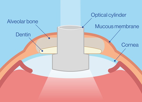
A pioneering idea: the principle of biointegration 💡
For decades, attempts at corneal prostheses have been thwarted by the body’s rejection. The stroke of genius of Italian surgeon Benedetto Strampelli in the 1960s was to use a part of the patient’s own body: a tooth (often a canine) with its supporting bone. This living support, recognised and nourished by the organism, allows for the stable and durable fixation of an optical lens, thus preventing rejection. This proven technique, now improved, is proof of its conceptual robustness.
OOKP: a last-resort procedure
Who may be eligible?
- Legal blindness in both eyes caused by damage to the ocular surface, such as severe burns or autoimmune disease.
- An essential requirement is a fully functioning retina and optic nerve.
- Good overall health and the presence of at least one healthy tooth (preferably a canine).
Who is not eligible?
- No perception of light (non-functional optic nerve or retina).
- Advanced, uncontrolled glaucoma or irreparable retinal detachment.
- Inability to commit to rigorous, lifelong medical follow-up.
A Complex Surgical Procedure
OOKP is a remarkable procedure that takes place in two main stages, separated by several months, and requires collaboration between an ophthalmologist and a maxillofacial surgeon. In France, Professor Vincent Daien recently performed this procedure on a young patient.
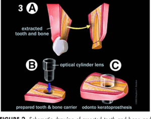
The surgeon removes a tooth and its bony support to insert an optical cylinder made of acrylic. The entire structure is then placed under the skin (for example, in the cheek) for 2 to 4 months. This crucial step allows the body to vascularise the graft, transforming it into a living implant ready to be transplanted into the eye.
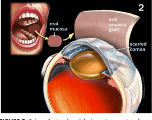
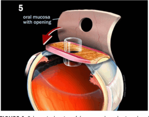
A few months later, the vascularised prosthesis is retrieved. The surgeon prepares the eye by creating an opening in the centre of the opaque cornea and inserting the prosthesis. The surface of the eye is covered with a membrane taken from the cheek, and only the optical lens protrudes. This new “window” allows light to finally reach the retina.
A New Window on the World
The restored vision can be spectacular, allowing patients to read, recognise faces and move independently. However, it is fundamental to understand that vision is not “normal.” The patient sees through a small cylinder, which results in tubular vision, comparable to that seen through a keyhole. Central vision is excellent, but peripheral vision is very limited, requiring training to “scan” the world by turning the head. The appearance of the eye is also altered, an important aspect to accept.
Risks and Follow-up
The success of OOKP depends on the patient’s unwavering commitment to lifelong follow-up to detect and treat complications.
Glaucoma: The Silent Enemy
This is the most feared long-term complication, affecting a significant number of patients. Its monitoring is complex because traditional instruments for measuring intraocular pressure are ineffective. Follow-up relies on imaging techniques (ultrasound) and palpation of the eyeball.
Other risks include retinal detachment, complications related to the prosthesis itself or the surface membrane, and, more rarely, serious infections.
References
À lire aussi
Your ophthalmologist answers your questions during the consultation. For a sudden symptom or sudden loss of vision, seek advice promptly: see the eye emergencies page.